Recognizing the Signs of Overuse Knee Pain and When to Act
Knee pain from overuse is a common issue affecting athletes, active individuals, and those with physically demanding jobs. It often develops gradually due to repetitive activities or insufficient recovery, leading to inflammation and tissue damage. Early recognition of symptoms and understanding the causes are essential steps in effective management. This article explores how to identify overuse knee injuries, differentiate them from other causes, and implement treatment and prevention strategies to maintain healthy knees.
Recognizing Symptoms of Overuse Knee Pain
How can you recognize symptoms of knee pain caused by overuse?
Knee pain stemming from overuse typically manifests as discomfort or ache concentrated in specific parts of the knee, such as the front, sides, or back. Recognizing these symptoms early is essential for proper management and to prevent further injury.
One of the most telling signs is pain that intensifies with activity. Activities like running, jumping, cycling, or even prolonged walking can trigger or exacerbate the discomfort. This type of pain often recurs after exercise or strenuous movement, signaling repetitive strain.
In addition to localized pain, individuals may experience other symptoms such as catching, locking, or a sensation of the knee giving way. Swelling and tenderness around the joint are common, and the warmth or redness might indicate inflammation.
Repetitive motions can cause inflammation in various soft tissues around the knee, including tendons, bursae, and the iliotibial band. For example,
- Tendinitis involves tendon inflammation, leading to pain at the front of the knee, especially during activities like jumping or stair climbing.
- Bursitis occurs when fluid-filled bursae become inflamed, causing swelling, tenderness, and pain.
- Iliotibial band syndrome results in pain on the outer side of the knee, commonly affecting runners, particularly after downhill running or frequent cycling.
Some overuse injuries have characteristic patterns. Patellofemoral Pain Syndrome, or 'runner’s knee,' typically causes pain behind or around the kneecap that worsens when extending the knee or sitting for long periods.
Early detection involves paying attention to how pain correlates with specific activities or movements. Noticing that the pain develops gradually, worsens with activity, and recedes with rest can help identify overuse as the culprit.
Assessment by a healthcare professional may include examining tender points, swelling, and joint stability, along with an activity history review. Imaging tests such as ultrasound or MRI can help visualize soft tissue inflammation or damage.
In summary, recognizing overuse knee pain involves observing the location, pattern, and triggers of pain, along with associated symptoms like swelling and stiffness. Addressing these signs promptly can prevent progression to more severe injuries and facilitate effective treatment.
Causes, Risk Factors, and Patterns of Overuse Knee Injuries
What are the causes and risk factors for overuse knee injuries?
Overuse injuries of the knee occur when repetitive loading and microtrauma damage the tendons, ligaments, cartilage, or bursa within the joint. These injuries develop gradually through activities that repeatedly stress the knee, such as jumping, running, cycling, or prolonged kneeling. When tissues are subjected to continuous strain without sufficient time for repair, inflammation and degeneration can ensue, leading to conditions like tendinitis, bursitis, or patellofemoral pain syndrome.
A primary cause is excessive or repetitive motion that exceeds the tissue’s capacity to recover. For instance, a runner who suddenly increases mileage or intensity may develop runner’s knee or iliotibial band (ITB) syndrome. Similarly, sports involving repeated jumping or sudden directional changes can overwhelm the knee’s stabilizing structures.
Several factors increase vulnerability to overuse injuries. Sudden spikes in activity levels or training volume without proper conditioning can overload the tissues. Poor biomechanics, such as misalignment or improper gait during exercise, can concentrate stress on certain parts of the knee.
Inadequate footwear or worn-out shoes also contribute, as they fail to provide sufficient support and shock absorption. Early sport specialization, where young athletes focus intensely on one activity, predisposes them to repetitive microtrauma because of lack of varied movement and insufficient rest.
Conditions like jumper’s knee (patellar tendinitis), IT band syndrome, bursitis, and osteoarthritis often arise from these repetitive stresses. Muscle imbalances—such as weak quadriceps or tight hamstrings—affect joint stability and increase strain on specific structures.
Environmental factors, including running on hard surfaces or uneven terrains, can further exacerbate stress on the knee. Overtraining without adequate rest impairs the tissue’s ability to recover, leading to accumulated damage.
Prevention strategies include proper warm-up routines, stretching, strengthening exercises for supporting muscles, using suitable footwear, and avoiding sudden increases in activity. Recognizing early signs and allowing sufficient recovery are essential to prevent progression to more severe injury.
In summary, overuse knee injuries are largely caused by repetitive microtrauma from physical activities and can be exacerbated by biomechanical faults, poor equipment, and training errors. Addressing these risk factors through balanced training, proper technique, and sufficient rest helps in safeguarding knee health and promoting recovery.
Differentiating Overuse Injury from Other Knee Lesions
How can you differentiate overuse knee pain from other types of knee injuries?
Understanding the differences between overuse injuries and traumatic knee lesions is essential for proper diagnosis and treatment. Overuse knee pain typically develops gradually over time, often associated with repetitive activities that strain the joint, such as running, cycling, or jumping.
In cases of overuse, symptoms tend to worsen with continued activity and may improve with rest. Patients often experience localized tenderness, mild swelling, and discomfort that does not immediately limit movement but can become persistent if not managed. Examples include conditions like patellar tendinitis (jumper’s knee) or iliotibial band syndrome, which cause specific, activity-related pain.
Conversely, traumatic injuries usually occur suddenly after an identifiable event like a twist, fall, or direct blow. Symptoms often include immediate severe pain, noticeable swelling, bruising, or deformity. Such injuries may involve ligament tears, meniscus injuries, fractures, or dislocations.
Signs such as locking or clicking are indicative of soft tissue damage, especially in meniscal tears or ligament injuries, rather than overuse problems. For instance, a knee that buckles or feels unstable may suggest ligament damage, particularly when accompanied by a popping sound during injury.
In traumatic cases, there is often difficulty bearing weight or straightening the knee fully. Swelling and bruising tend to appear rapidly. In contrast, overuse injuries may show a gradual increase in discomfort without significant acute swelling or deformity.
Recognizing these differences allows for accurate diagnosis. Persistent or worsening knee pain, especially if coupled with swelling, deformity, or instability, warrants prompt medical evaluation. A healthcare provider can then perform the necessary physical examination and imaging tests, such as X-rays or MRI, to identify the exact injury and determine the best course of treatment.
Diagnostic Methods for Knee Pain Evaluation
What are the methods for diagnosing and evaluating knee pain?
Diagnosing the cause of knee pain involves a systematic approach combining history-taking, physical examination, and various diagnostic tests. The process aims to identify whether the pain stems from injuries, arthritis, overuse, or other underlying conditions.
Patient history is an essential first step. The healthcare provider considers the patient's activity levels, recent injuries, onset and nature of pain (sudden or gradual), and factors that worsen or relieve symptoms. Understanding the patient's occupation, sports participation, and weight status helps identify potential risk factors for specific conditions.
Physical examination is vital for assessing the knee. The clinician inspects for swelling, tenderness, warmth, skin discoloration, and signs of deformity. They also evaluate the range of motion, strength, and stability of the knee joint. Special tests may be performed to detect ligament injuries (like ACL or MCL sprains), meniscus tears, or patellofemoral instability. The examiner may also check for signs of infection or bursitis.
Imaging studies are crucial tools in the evaluation process. Commonly used imaging modalities include:
| Imaging Test | Purpose | Additional Details |
|---|---|---|
| X-ray | Detects fractures, osteoarthritis, bone spurs | Quick, widely available, no soft tissue detail |
| MRI | Visualizes soft tissues, ligaments, menisci | High-resolution, essential for soft tissue injuries |
| Ultrasound | Guides injections, detects bursitis, tendinitis | Dynamic assessment during movement |
| CT Scan | Detailed bone imaging, complex fractures | Often used if X-ray is inconclusive |
| Radionuclide scans | Detects bone infections, tumors | Less common, more specialized imaging |
Laboratory tests may be performed if infection, autoimmune disease, or inflammation is suspected. These include blood tests such as ESR, CRP, rheumatoid factor, and uric acid levels. Arthrocentesis, which involves aspirating fluid from the joint, can help diagnose infection or gout.
Arthroscopy is a minimally invasive surgical procedure that provides direct visualization of internal joint structures. During arthroscopy, surgeons can inspect, diagnose, and treat issues such as meniscus tears or loose bodies, offering a definitive diagnosis and therapeutic intervention.
Summary:
| Diagnostic Method | Main Use | Benefits | Limitations |
|---|---|---|---|
| Patient History | Understanding symptoms and cause | Guides focus for exams and tests | Subjective, relies on patient recall |
| Physical Exam | Detecting physical signs | Immediate, bedside assessment | May not visualize internal issues |
| X-ray | Bone abnormalities | Fast, accessible | No soft tissue detail |
| MRI | Soft tissue injuries | Detailed, comprehensive | Costly, less available |
| Ultrasound | Bursitis, tendinitis, guiding injections | Dynamic, real-time imaging | Operator-dependent |
| CT Scan | Complex bone injuries | High detail for bones | Higher radiation exposure |
| Blood Tests | Inflammatory, autoimmune, infectious conditions | Helps diagnose systemic causes | Not specific for structural injuries |
| Arthroscopy | Internal joint visualization | Direct diagnosis and treatment | Invasive, requires anesthesia |
Effective diagnosis of knee pain often requires integrating findings from history, physical exam, imaging, and laboratory tests. This comprehensive assessment guides targeted treatment plans, whether conservative or surgical.
Search term for further research:
Methods for diagnosing knee overuse injuries
Treatment Strategies for Overuse Knee Injuries
What treatment options are available for overuse knee injuries?
Managing overuse knee injuries involves a combination of approaches aimed at easing symptoms, promoting healing, and preventing future issues. The initial step typically includes conservative measures such as the RICE protocol—rest, ice, compression, and elevation. Rest is critical to allow inflamed tissues to recover, while applying ice helps reduce swelling and inflammation. Compression with bandages or braces provides support and decreases movement that could aggravate the injury. Elevation keeps fluids from pooling around the knee, alleviating swelling.
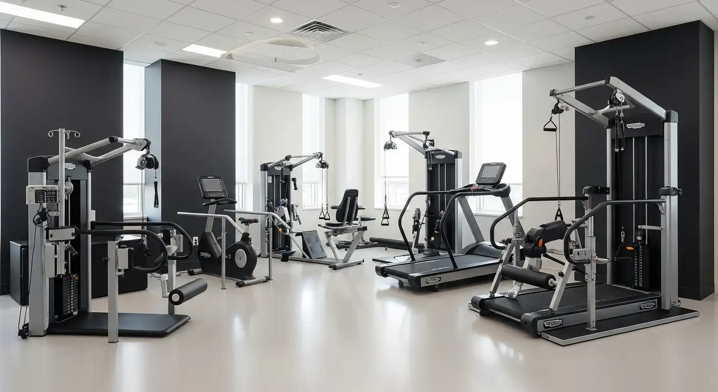
Physical therapy is often central to treatment, focusing on gentle stretching and strengthening exercises. These exercises improve flexibility, correct biomechanical issues, and rebuild muscle support around the joint, which is essential in conditions like jumper’s knee or iliotibial band syndrome.
Medications are also recommended to manage pain and inflammation. Non-steroidal anti-inflammatory drugs (NSAIDs) like ibuprofen or naproxen can be effective, along with acetaminophen for pain relief if inflammation isn't a major concern.
Supportive devices such as knee braces, taping, or custom orthotics may be used to stabilize the knee, offload stress, and facilitate proper alignment during activity.
When conservative treatments do not yield sufficient relief, more advanced options are considered. Corticosteroid injections can provide rapid reduction of inflammation and pain in specific cases, especially for bursitis or severe tendinitis.
Surgical interventions, including arthroscopy, might be necessary for repairing torn tissues, removing inflamed bursae, or correcting structural abnormalities. Surgery aims to restore joint function, reduce pain, and prevent further deterioration.
Early diagnosis and a structured, multimodal treatment plan are critical for optimal outcomes. This comprehensive approach helps prevent the progression to chronic problems, facilitates full recovery, and returns individuals to their normal activities and sports.
| Treatment Area | Techniques & Measures | Purpose & Details |
|---|---|---|
| Initial management | Rest, ice, compression, elevation (RICE) | Reduce swelling, control inflammation, provide peace to the joint |
| Physical therapy | Stretching, strengthening exercises, biomechanical correction | Enhance flexibility, rebuild support muscles, correct movement patterns |
| Medications | NSAIDs, acetaminophen | Pain relief, inflammation reduction |
| Supportive devices | Braces, taping, orthotics | Stability, support, proper alignment |
| Advanced therapies | Corticosteroid injections, surgery (arthroscopy, repair) | Targeted intervention for severe or persistent cases |
Understanding and applying these treatment options can significantly improve recovery chances, diminish symptoms, and restore knee function after overuse injuries.
Home Care and Preventive Strategies
What are effective home care and self-management strategies for knee pain?
Managing knee pain at home involves several proven methods to alleviate discomfort and prevent worsening of conditions. One fundamental approach is the RICE method—Rest, Ice, Compression, and Elevation. Resting the knee helps reduce inflammation and prevents further injury. Applying ice packs during the first 48 hours after pain onset helps diminish swelling and dulls pain signals. Compression with an elastic bandage or knee sleeve stabilizes the joint, while elevating the leg above heart level promotes fluid drainage, further reducing swelling.
In addition to RICE, heat therapy can be useful for relaxing tense muscles and increasing blood flow once initial inflammation subsides. Gentle stretching exercises targeting the hamstrings, quadriceps, and calf muscles improve flexibility and reduce tension around the knee joint. Strengthening exercises focusing on the muscles supporting the knee—such as leg presses, wall sits, and targeted physiotherapy routines—enhance joint stability and decrease strain.
Weight management plays a crucial role in knee health. Excess weight increases pressure on the knees, magnifying wear and tear. Maintaining a healthy weight through diet and appropriate exercise reduces stress, alleviating pain and improving function.
Proper footwear is also vital. Shoes with good cushioning and stability help absorb shock during activities and prevent abnormal joint movements that could lead to injuries. Replacing worn-out shoes and considering orthotics, especially for flat feet or other structural issues, can substantially benefit knee health.
Avoiding high-impact activities such as running, jumping, or heavy lifting during pain episodes prevents worsening symptoms. Instead, low-impact options like swimming, cycling, or elliptical training provide cardiovascular benefits while minimizing knee stress.
Monitoring symptoms is essential. If pain persists beyond a few days, worsens, or if there are signs of swelling, redness, warmth, or instability, it is advisable to seek medical attention. A healthcare professional can perform further assessments, recommend imaging tests, or suggest advanced treatments such as physical therapy, injections, or surgery if necessary.
In summary, effective self-care for knee overuse injuries combines the RICE approach, targeted exercises, weight control, proper footwear, activity modification, and vigilant symptom monitoring—forming a comprehensive strategy to maintain knee health and prevent chronic issues.
Additional tips for knee health include:
- Warming up thoroughly before exercise.
- Cooling down and stretching post-activity.
- Gradually increasing exercise intensity.
- Using supportive braces if recommended.
- Listening to your body and avoiding overexertion.
Applying these principles consistently can significantly improve knee comfort and function, helping individuals stay active and reduce the likelihood of future problems.
Searching for more information:
For detailed self-care practices and injury-specific advice, search terms such as "Self-care for knee overuse injuries" can provide targeted guidance and professional recommendations.
Long-term Management and When to Seek Help
Persistent or severe knee pain from overuse should not be ignored. Long-term management involves ongoing strength training, proper biomechanics, weight control, and activity moderation. Recognizing early symptoms and implementing preventative measures can reduce the risk of chronic damage and preserve knee health. Consultation with healthcare providers is essential if pain worsens, if there are signs of instability, or if initial home treatments do not provide relief within a few days. With timely action and comprehensive care, overuse knee injuries can be effectively managed, helping maintain active, pain-free lifestyles.
References
- Knee Pain: Causes & Treatment - Cleveland Clinic
- Knee Pain - Symptoms and Causes - Penn Medicine
- Overuse of Knee Injuries & Conditions - The Yorkshire Knee Clinic
- Patient education: Knee pain (Beyond the Basics) - UpToDate
- Overuse Injuries | Johns Hopkins Medicine
- Types and Causes of Common Knee Injuries and Problems
- Common knee injuries and how you can avoid them
- Knee pain: MedlinePlus Medical Encyclopedia
- How to Get Rid of Knee Pain & When to See a Doctor | Houston ...
- Leg & Knee Overuse Injuries - Inspira Health Network