Comprehensive Insights into Lateral Posterior Knee Pain
Lateral posterior knee pain is a significant complaint among patients presenting with knee discomfort, often associated with complex injuries to the posterolateral corner (PLC) of the knee. This article explores the anatomy, injury mechanisms, diagnostic strategies, and treatment options for lateral posterior knee pain, aiming to equip clinicians and patients with an in-depth understanding of this challenging condition.
Anatomy of the Knee and the Posterolateral Corner
Basic knee anatomy including bones, ligaments, tendons, muscles, and bursa
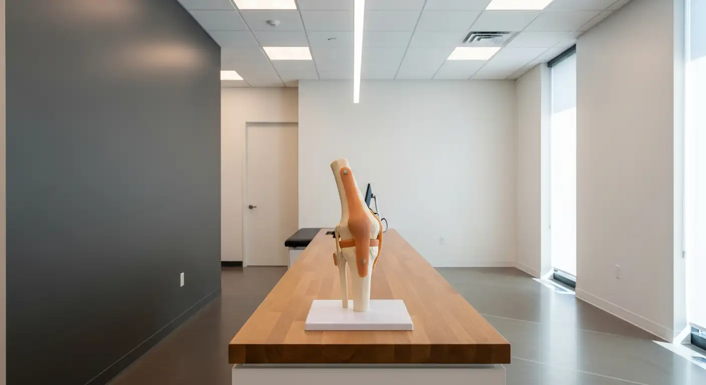
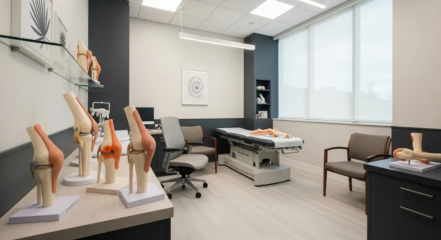
The knee is a complex joint comprising several bones, soft tissues, and cartilage. Its primary bones include the femur (thigh bone), tibia (shin bone), and patella (kneecap). The articular surfaces are covered with hyaline cartilage, facilitating smooth movement. The menisci—medial and lateral—are cartilaginous discs acting as shock absorbers.
Ligaments provide stability, notably the anterior cruciate ligament (ACL), posterior cruciate ligament (PCL), medial collateral ligament (MCL), and lateral collateral ligament (LCL). Tendons connect muscles to bones; major tendons include those of the hamstrings, gastrocnemius, and quadriceps.
Muscles such as the hamstrings, gastrocnemius, and popliteus play vital roles in knee movement and stability. The knee also contains bursae—fluid-filled sacs—that reduce friction during motion.
Structures involved in the posterolateral corner such as the lateral collateral ligament, popliteus tendon, popliteofibular ligament, and associated musculature
The posterolateral corner (PLC) is a complex anatomical region on the outside part of the knee. Key structures include the lateral collateral ligament (LCL), popliteus tendon, popliteofibular ligament, and the arcuate ligament.
The LCL runs from the lateral femoral epicondyle to the fibular head, resisting varus stress. The popliteus tendon originates from the lateral femoral condyle, wrapping around the posterior lateral aspect, attaching to the tibia, and acting to internally rotate and unlock the knee.
Additional structures such as the biceps femoris tendon, the fibular head, and the lateral head of the gastrocnemius also contribute to the stability of the lateral knee. The peroneal nerve, which wraps around the fibular neck, is closely associated with the posterolateral corner and vulnerable in injuries.
Functions of the PLC in knee stability, particularly resisting external rotation and posterior translation of the tibia
The PLC plays a crucial role in maintaining posterior and rotational stability of the knee. It primarily acts to resist excessive external rotation of the tibia, especially during movements such as twisting or pivoting. It also prevents posterior translation—backward slipping—of the tibia relative to the femur.
This stability is vital in activities involving sudden changes in direction or lateral forces, common in sports and accidents. Damage to the PLC can compromise knee stability, leading to instability, increased risk of further injuries, and long-term degenerative changes.
Understanding the detailed anatomy and function of the posterolateral corner helps in diagnosing injuries and planning effective surgical or conservative treatment strategies.
| Structure | Function | Involved in Stability | Common Injury Type |
|---|---|---|---|
| Lateral collateral ligament (LCL) | Resists varus stress and external rotation | Lateral stability, resisting outward bending | Sprain, tear |
| Popliteus tendon | Unlocks the knee from extension, internal rotation | Internal rotation, posterior stability | Tendinopathy, tear |
| Popliteofibular ligament | Stabilizes posterolateral corner | Limits external rotation and posterior translation | Partial or complete tear |
| Biceps femoris tendon | Assists in knee flexion and stabilization | Lateral stability, knee flexion | Strain, rupture |
| Fibular head | Point of attachment for ligaments and muscles | Supports LCL and stability | Fracture, ligament injury |
| Peroneal nerve | Innervates the lateral lower leg and foot | Sensory and motor function, risk in injuries | Nerve injury, foot drop |
By comprehensively understanding these tissues, clinicians can better identify the cause of lateral or posterolateral knee pain and tailor effective treatments.
Key Takeaways and Future Directions
Lateral posterior knee pain encompasses a spectrum of conditions rooted in the complex anatomy and biomechanics of the posterior and lateral aspects of the knee. Accurate diagnosis hinges on a detailed clinical examination and advanced imaging modalities like MRI, which reveal the extent of soft tissue damage. Recognizing the signs and symptoms associated with PLC injuries, such as instability, pain during twisting or pivoting, and nerve involvement, is critical for timely and appropriate management. Treatment strategies range from conservative approaches, including physical therapy and bracing, to surgical reconstruction for severe injuries. Emphasizing early diagnosis and intervention can prevent long-term complications such as chronic instability, osteoarthritis, and graft failure. Ongoing research into reconstruction techniques and biologic therapies promises to enhance treatment outcomes, offering hope for patients with complex lateral posterior knee pain.
References
- Posterolateral Corner (PLC) Knee Injuries
- Posterolateral Knee Injuries - Robert LaPrade, MD
- Posterior Knee Pain
- Posterior knee pain - PMC
- Pain behind the Knee (posterior pain): Causes and treatment
- Posterolateral Corner Injuries: Everything You Need To Know
- Posterolateral corner knee injuries: a narrative review - PMC
- Knee Pain in Adults and Adolescents: The Initial Evaluation