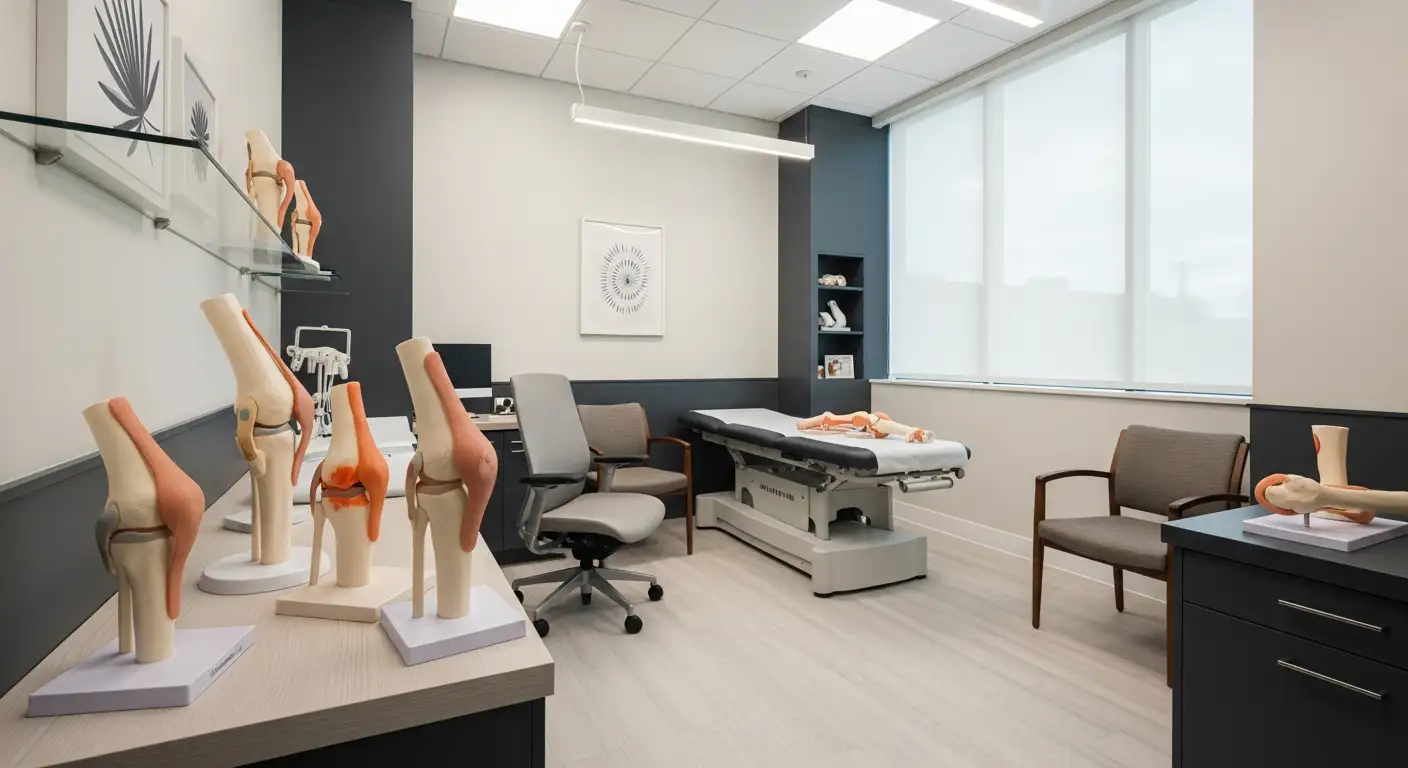
Decoding the Complexities of Lock Knee
Lock Knee is a condition characterized by the inability to fully bend or straighten the knee joint, often accompanied by pain, swelling, and a feeling of the knee being stuck. It can result from various intra-articular or extra-articular issues, affecting individuals of all ages and activity levels. This comprehensive overview delves into the causes, symptoms, diagnostic methods, and treatment strategies for Lock Knee, helping readers understand this condition's complexities and management options.
Symptoms of Lock Knee and Their Clinical Significance
What are the common symptoms of a locked knee?
A locked knee typically presents with difficulty in fully bending or straightening the joint. Patients often report an inability to move the knee freely, which can interfere with daily activities. Accompanying these movement restrictions, pain is a prevalent symptom, especially during attempts to bend or straighten.
Swelling around the knee is also common, indicating inflammation or internal injury. Many individuals describe a sensation that their knee is stuck or catches during movement, which can be startling and painful.
Another hallmark feature is the popping or clicking sound during joint motion. These noises often occur when there is displacement of tissues like torn cartilage or loose bodies in the joint.
In cases of true locked knee, a visible bump or loose piece of tissue such as a torn meniscus or fragment of cartilage can sometimes be felt or seen in the joint. Stiffness is frequently reported, making it difficult to extend the knee completely.
Pseudo locked knee differs as it is mainly caused by pain-induced muscle spasms rather than an actual physical obstacle, leading to sensations of catching or instability without a mechanical block. Conversely, true locked knee involves a mechanical barrier, such as a torn meniscus or loose cartilage fragment, physically preventing movement.
Overall, these symptoms often become apparent during activities that require knee movement or weight bearing. Many cases follow trauma or injury, making early diagnosis and treatment critical to maintaining joint function.
Underlying Causes and Risk Factors for Lock Knee
 The causes behind a locked knee primarily revolve around structural and functional abnormalities within the joint. Structural issues such as meniscal tears, loose bodies, ligament injuries, and osteoarthritis are common culprits. Meniscal tears, especially bucket handle types, can physically obstruct movement if a fragment slips into the joint space, preventing full extension. Loose bodies—small fragments of cartilage or bone—can also float within the joint and cause mechanical blockage.
Ligament injuries, notably anterior cruciate ligament (ACL) tears, may lead to instability and locking when torn tissue catches between joint surfaces. Severe degenerative conditions like osteoarthritis cause cartilage breakdown, bone spurs, and loose fragments that can mechanically hinder movement.
Pseudo locking, on the other hand, is caused by pain and muscle spasms rather than a physical obstacle. It occurs when severe pain from injury, inflammation, or other conditions triggers muscles to contract involuntarily, creating a sensation of the knee being stuck.
Various risk factors contribute to the development of a locked knee. Traumatic injuries from falls, sports, or accidents are common triggers, especially twisting injuries that damage menisci or ligaments. Degenerative joint diseases such as osteoarthritis increase the likelihood over time due to wear and tear on joint components.
Inflammatory diseases including rheumatoid arthritis or gout can cause swelling and deposits within the joint, further facilitating locking symptoms. Repetitive stress or overuse can weaken structures and predispose individuals to injury.
Patellar maltracking, where the kneecap shifts out of its normal path, can induce episodes of locking and instability. Conditions like bursitis, plica syndrome, and dislocated kneecaps also contribute to joint locking by interfering with movement or causing pain-induced muscle spasms.
In summary, a variety of structural injuries and degenerative conditions, combined with trauma and inflammation, render the knee susceptible to locking episodes. Early diagnosis and understanding of these risk factors are vital for effective treatment and prevention.
Diagnosis and Differentiation of Lock Knee from Other Conditions
How do you diagnose a locked knee and distinguish it from other knee conditions?
Diagnosing a locked knee involves a careful combination of patient history, physical examination, and imaging studies. The healthcare provider begins by asking about recent trauma, the onset of symptoms, and specific sensations such as catching, locking, or clicking. Physical examination focuses on observing the knee’s range of motion, noting any inability to fully bend or straighten the joint, presence of swelling, tenderness, or joint warmth.
During the exam, the clinician assesses for signs of internal joint obstruction, such as a palpable bump or laxity that may suggest a meniscal tear or loose body. The sensation of catching or locking may indicate a mechanical block like a torn meniscus, whereas swelling and pain without mechanical blockage might suggest inflammatory causes like arthritis.
Distinguishing between pseudo-locked and true locked knee is crucial:
- Pseudo locked knee is caused by muscle spasms or pain-induced restrictions. It often improves with rest, anti-inflammatory medications, or muscle relaxation.
- True locked knee involves an actual physical obstacle inside the joint, such as a displaced meniscal fragment, loose bone or cartilage, or ligament remnants preventing movement.
Imaging plays an essential role:
| Imaging Modality | Purpose | Typical Findings |
|---|---|---|
| X-ray | Detect bone fractures, osteoarthritic changes, loose bodies | Bone spurs, fracture lines, joint space narrowing |
| MRI | Visualize soft tissue structures like menisci, ligaments, and cartilage | Meniscal tears, ligament ruptures, intra-articular debris |
| CT Scan | Better visualization of complex bone fractures or calcifications | Bone fragments, detailed bony anatomy |
| Ultrasound | Assess soft tissues and effusion | Synovitis, joint capsule swelling |
In cases where imaging results are inconclusive, diagnostic arthroscopy is considered the gold standard. It involves a minimally invasive procedure where a small camera allows direct visualization of intra-articular structures. Arthroscopy not only confirms the diagnosis but also allows immediate treatment—such as removing loose bodies or repairing torn menisci.
Overall, accurate diagnosis relies on correlating clinical findings with imaging results, and in uncertain cases, arthroscopy provides definitive internal examination. Identifying the exact cause—whether mechanical obstruction or muscular spasm—guides suitable treatment and improves outcomes.
Treatment and Management of Lock Knee: From Conservative to Surgical Approaches
What are the treatment options and management strategies for a locked knee?
Treatment for a locked knee depends on its cause, whether it is true locking due to a physical obstacle or pseudo locking caused by pain and muscle spasms.
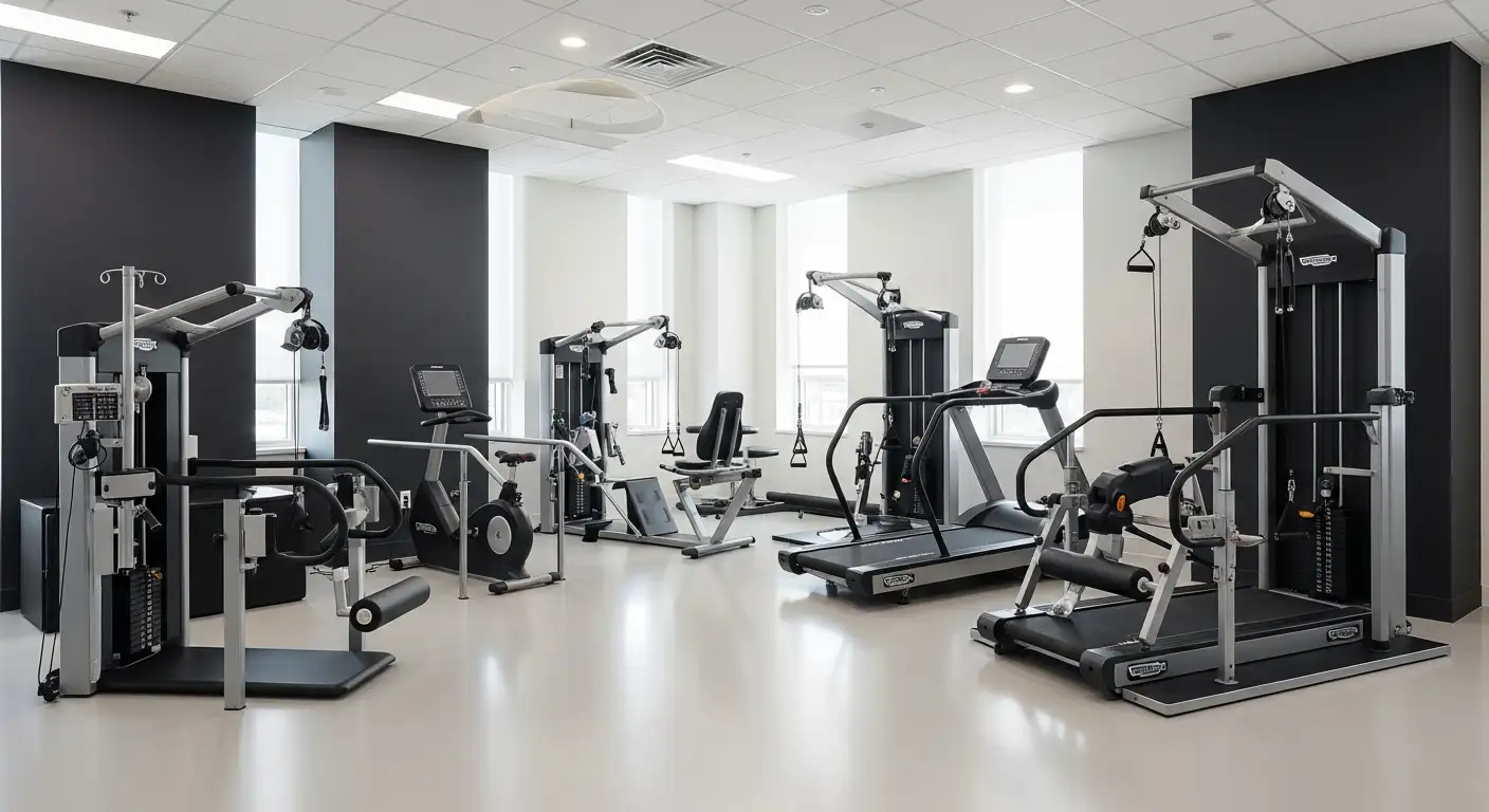
For true mechanical lockings, management usually begins with conservative measures like rest, ice, compression, elevation (RICE), alongside NSAIDs to reduce inflammation and pain. Physical therapy exercises play an important role in strengthening muscles around the knee, enhancing stability, and improving flexibility. Gentle exercises such as knee extensions, hamstring curls, and stretching routines are often recommended.
If symptoms persist despite conservative treatment, minimally invasive surgical procedures like arthroscopy are considered. During arthroscopy, damaged tissue—such as torn menisci, loose bodies, or cartilage fragments—can be removed or repaired, restoring knee function.
In cases where the locking is caused by specific conditions like gout, management includes medications to lower uric acid levels and anti-inflammatory drugs to control flare-ups. For loose bodies or displaced cartilage fragments causing obstruction, surgical removal via arthroscopy provides relief.
Ligament injuries, such as ACL tears, may require surgeries like ligament reconstruction to stabilize the joint. For patellar dislocations, realignment procedures or bracing can help prevent recurrence.
Post-treatment, physiotherapy continues to be vital. Rehabilitation focuses on restoring full range of motion, strengthening muscles, and preventing future episodes.
Deciding between surgical and conservative management depends on the severity of the lock, underlying cause, and response to initial treatments. Early consultation with an orthopedic specialist ensures the appropriate intervention to regain mobility and reduce symptoms effectively.
Home Remedies and Physiotherapy Techniques for Symptom Relief
 Managing a locked knee often involves simple home remedies and gentle physiotherapy techniques aimed at reducing pain, inflammation, and improving joint mobility. One of the most effective initial steps is following the RICE method—rest, ice, compression, and elevation. Resting the knee minimizes stress on the joint, while applying ice packs helps decrease inflammation and numbness, providing pain relief. Compression with an elastic bandage offers support and reduces swelling, and elevating the leg above heart level further diminishes fluid accumulation.
In addition to RICE, gentle stretching and knee exercises can be beneficial, especially for pseudo-locked knee caused by muscle spasms. Exercises like hamstring curls, knee extensions, and gentle knee bends aid in restoring flexibility and preventing stiffness. Regular movement helps maintain joint function and can alleviate symptoms. Heat therapy, such as warm packs or warm baths, relaxes tense muscles and increases blood flow, which can be soothing. Conversely, cold therapy reduces swelling and numbness, making it useful after activity.
Over-the-counter pain medications, including NSAIDs (nonsteroidal anti-inflammatory drugs) like ibuprofen and topical analgesic creams, can provide additional relief from pain and inflammation. Engaging in low-impact activities such as swimming and walking enables exercise without stressing the knee joint. Maintaining a healthy weight decreases undue pressure on the knee, reducing the risk of locking episodes.
Preventive measures are crucial for overall knee health. Warming up before exercise, wearing proper footwear, and strengthening muscles around the knee—including quadriceps and hamstrings—help support the joint. Proper technique and avoiding high-impact or twisting activities can further prevent future episodes of locking.
While these home remedies and physiotherapy techniques are effective for mild cases, persistent or severe symptoms warrant consultation with a healthcare professional to rule out underlying conditions needing specialized treatment. Early intervention can enhance recovery and prevent long-term damage.
Can a Locked Knee Resolve Naturally or Require Medical Intervention?
Can a locked knee resolve on its own, or does it require medical intervention?
A locked knee may or may not resolve without treatment, depending on what causes it. Pseudo locked knees, which happen due to muscle spasms or pain, often improve with conservative care. Rest, ice, anti-inflammatory medications, and physical therapy can help relax muscles and reduce swelling, leading to natural improvement.
On the other hand, true locked knees are caused by physical obstructions like torn menisci, loose bodies, or cartilage fragments. These issues generally do not get better on their own and often need medical treatments such as arthroscopic surgery to remove or repair damaged tissues.
If a lock persists or worsens, it’s important to see a healthcare professional promptly. Ignoring the problem can lead to joint damage, increased pain, and loss of knee function. Therefore, minor cases might resolve naturally, but severe or ongoing locking requires Medical diagnosis and appropriate treatment to prevent further complications.
Prevention Strategies and Lifestyle Tips to Reduce Lock Knee Incidents
How can you prevent knee locking from happening?
Prevention of knee locking primarily involves taking care of your knee health and avoiding injury triggers. Strengthening the muscles surrounding the knee, such as the quadriceps, hamstrings, and calves, can improve joint stability and reduce instability that leads to locking episodes.
In addition to strengthening, regular stretching and warming up before physical activities help prepare the knee for movement, lowering the risk of sudden injuries like meniscus tears or ligament injuries.
Being mindful during activities also plays a role. Avoid high-impact sports or activities that put excessive stress on the knees, especially if you already have joint problems like osteoarthritis. Wearing supportive footwear is essential, as proper shoes can absorb shock and reduce joint stress during walking, running, or other weight-bearing exercises.
Managing pre-existing conditions like arthritis is crucial. Proper medication, physical therapy, or lifestyle adjustments can slow joint deterioration and prevent incidental locking.
Furthermore, avoid sudden twisting, over-flexing, or hyperextending movements that could injure the meniscus or ligaments. Listen to your body and stop activity if you experience pain or instability.
While not all causes are preventable, maintaining overall knee health, building muscle resilience, and exercising caution during physical activity significantly lower the chances of knee locking episodes. Early intervention at signs of discomfort can help prevent minor issues from developing into severe locking problems.
Summary and Final Thoughts on Lock Knee
Lock Knee is a multifaceted orthopedic condition that can significantly impact mobility and quality of life. Understanding its symptoms, causes, and treatment options enables timely intervention, which is crucial for optimal recovery. Whether caused by structural damage like meniscal tears or by muscle spasms in pseudo locking, early diagnosis using physical exams and imaging is vital. Management ranges from conservative approaches such as physiotherapy and NSAIDs to surgical procedures like arthroscopy, tailored to the underlying pathology. Preventive strategies focusing on muscle strengthening, flexibility, and avoiding injury are essential to reduce recurrence. If persistent or severe symptoms occur, seeking prompt medical attention can prevent further joint damage and ensure effective treatment, ultimately helping individuals regain joint function and prevent future episodes.
References
- Locked knee: Causes, symptoms, and treatment
- 7 Main Causes of Locked Knees | OrthoNeuro | Columbus, OH
- The unusual traumatic locked young knee - PMC
- Knee Popping, Locking, Buckling, and Giving Out
- Locked Knee: Causes, Treatments and When to Seek Help
- What causes a locked knee? - Chris Bailey Orthopaedics
- Knee Locking: Causes and Treatments for Relief - Hinge Health
- The 'Chalky Culprit' of acute locked knee - PMC