Understanding Snapping Biceps Femoris Tendon: Anatomy, Diagnosis, and Treatment
Snapping biceps femoris tendon is a rare but noteworthy cause of lateral knee pain characterized by a characteristic snapping or cracking sensation during knee movement. This condition often challenges clinicians due to its subtle presentation and complex anatomical underpinnings. This article aims to elucidate the anatomy involved, symptoms experienced, diagnostic approaches, causative factors, and the latest management strategies based on current literature and case studies.
Anatomical Foundations and the Pathophysiology of Snapping
What is the anatomy of the biceps femoris tendon and how does it relate to snapping phenomena?
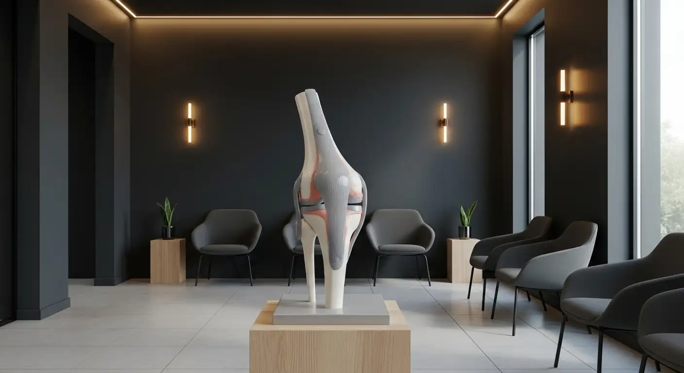
The biceps femoris tendon plays a vital role in knee stability and external rotation of the tibia. Its complex structure involves multiple slips, primarily attaching to the fibular head and occasionally extending to the lateral tibia. This intricate insertion pattern allows for versatile movement but also predisposes it to various abnormalities.
Variations such as anomalous insertions, where the tendon attaches in an atypical manner, or morphological irregularities of the fibular head, can lead to instability. These anatomical differences sometimes result in the tendon slipping or dislocating over the fibular head during knee motion.
This displacement—known as subluxation or dislocation—can cause a snapping sensation or sound, especially during deep knee flexion activities like squatting or sitting. Patients often report lateral knee pain accompanied by a visible or audible click.
Imaging techniques, particularly MRI, are instrumental in visualizing these anomalies. MRI can reveal thickening of the biceps femoris tendons, tears, or abnormal insertions, helping clinicians pinpoint the source of snapping.
Understanding the anatomy and its variations informs treatment strategies. Surgical options often involve reattaching or stabilizing the tendons, or smoothing bony features like fibular head prominences, to correct the motion and relieve symptoms.
Below is a summary table of the key anatomical features and their link to snapping phenomena:
| Anatomical Feature | Description | Implication in Snapping |
|---|---|---|
| Tendon slips | Multiple slips attaching to fibula and tibia | Variations can cause abnormal movement |
| Fibular head | Bony prominence where tendons insert | Morphological anomalies may contribute to tendon subluxation |
| Tendon insertions | Normal or anomalous attachments | Abnormal insertions increase risk of dislocation and snapping |
By examining these structures through imaging and physical assessment, clinicians can tailor interventions that restore normal movement and eliminate the snapping.
For further understanding, search: "biceps femoris tendon anatomy and snapping mechanisms." Overall, recognizing the detailed anatomy of this tendon is essential in diagnosing and managing snapping biceps femoris syndrome effectively.
Clinical Manifestations and Typical Presentation

What are the clinical signs and symptoms associated with snapping biceps femoris tendon?
Patients suffering from snapping biceps femoris tendon usually experience lateral knee pain accompanied by a distinctive snapping or clicking sensation over the outer knee region. These symptoms tend to worsen during activities that require deep knee bending, such as squatting, cycling, or climbing stairs.
During physical examination, clinicians may observe or feel a palpable subluxation of the distal biceps femoris tendon over the fibular head when the patient moves the knee through flexion and extension. This movement often reproduces the symptoms of snapping, confirming the clinical suspicion.
Pain is typically concentrated along the lateral aspect of the knee, particularly over the site where the biceps femoris tendon attaches. Tenderness may be present at the fibular head, highlighting the area of pathology.
In some cases, patients also report hearing a "pop" or "clunk" during knee motion, especially in deep flexion. Additionally, if the condition irritates the nearby common peroneal nerve, symptoms such as numbness or tingling sensations may occur in the lower leg.
Physical tests that involve pressing on the fibular head or moving the knee into flexion and extension can help reproduce the snapping phenomenon. Imaging studies, like ultrasound or MRI, further aid in confirming the diagnosis and ruling out other issues such as ligament tears or meniscal injuries.
Overall, these clinical signs and symptoms are pivotal for diagnosing snapping biceps femoris tendon and determining appropriate treatment strategies.
Diagnostic Strategies: Clinical and Radiographic Insights

How is snapping biceps femoris tendon diagnosed clinically and radiographically?
Diagnosing a snapping biceps femoris tendon requires a comprehensive approach that includes both detailed patient history and thorough physical examination. Patients often report lateral knee pain coupled with a noticeable or audible snapping sensation during deep knee flexion activities like squatting or sitting low. They may describe episodes of the tendon subluxating over the fibular head.
During physical assessment, clinicians look for signs of subluxation or snapping by palpating the lateral knee while the patient moves through various ranges of motion. Manual pressure over the fibular head may reduce or reproduce the snapping, which helps confirm the diagnosis. Additional examinations include evaluating knee stability, checking for tenderness, assessing range of motion, and ruling out other pathologies such as meniscal tears or ligament injuries.
Imaging modalities play an essential role in visualizing both soft tissue and bony structures. Ultrasound is particularly useful for real-time, dynamic assessment of the tendon during movement. It can reveal thickening of the biceps femoris tendon, degenerative changes, or abnormal fibular head prominences that contribute to snapping. MRI offers a detailed view at the soft tissue level, showing tendon thickening, degenerative changes, partial tears, or detachments of the biceps femoris tendons. MRI can also uncover bony abnormalities like fibular head prominence or morphological variations in tendon insertion.
While plain radiographs may not show soft tissue issues, they are valuable for detecting bony abnormalities, such as an enlarged or prominent fibular head, which could predispose to snapping. These bony features can often be correlated with clinical findings.
In practice, diagnosis involves integrating history, physical examination findings, and imaging results. When necessary, intraoperative observations during surgical repair can further confirm the diagnosis, especially in complex or recurrent cases. This combined approach ensures an accurate diagnosis and guides effective treatment, whether conservative or surgical.
| Diagnostic Procedure | Focus Area | Useful Findings | Additional Notes |
|---|---|---|---|
| Patient history | Symptoms, activity triggers | Snapping, pain duration | Reinforces clinical suspicion |
| Physical exam | Palpation during movement | Subluxation, tenderness | Can reproduce snapping |
| Ultrasound | Tendon dynamic visualization | Tendon thickening, subluxation | Detect degenerative changes |
| MRI | Soft tissue details | Tendon degeneration, insertional abnormalities | Evaluate adjacent structures |
| Radiographs | Bone morphology | Fibular head positioning | Identify bony contributions |
Ultimately, diagnosis is a synthesis of clinical insights and imaging evidence, ensuring targeted treatment approaches.
Etiology: Causes and Predisposing Factors
 The snapping of the biceps femoris tendon is a rare but notable cause of lateral knee pain, often identified by a painful cracking sound during movement. Its development can be attributed to a range of anatomical and biomechanical factors.
One major cause involves anatomical variations, specifically anomalous tendon insertions. These include abnormal distal insertions of the long and short heads of the biceps femoris, such as excessive anterior placement on the fibular head or atypical tibial attachments. Such variations may predispose the tendon to subluxation or snapping over the fibular head.
Trauma or alterations in fibular head morphology also play significant roles. For example, prominence or thickening of the fibular head can create a rough surface that facilitates tendinous friction. The case report highlighted bilateral snapping caused by prominent fibular head features coupled with thickening of the anterior band of the biceps femoris tendons.
Repetitive knee flexion activities, including cycling, football, running, and deep squatting, increase stress on the lateral knee structures. These activities may exacerbate friction or cause tendinous subluxation over the fibular head, especially in the presence of anatomical abnormalities.
Patients with congenital or acquired deformities of the fibula or distal biceps femoris tendons are at heightened risk. The combination of structural variations and repetitive movement stressors fosters conditions conducive to snapping phenomena.
In summary, snapping biceps femoris tendon results from a combination of congenital anatomical variations—such as anomalous tendon insertions—and acquired factors like fibular head irregularities, aggravated by activity-related stresses. Recognizing these predisposing factors is essential for accurate diagnosis and appropriate management of this uncommon knee pathology.
Management Approaches: Conservative and Surgical Options

What treatment options are available for managing snapping biceps femoris tendon?
Treating snapping biceps femoris tendon involves a variety of approaches, depending on the severity of symptoms and the underlying anatomical issues. Initially, conservative methods are often attempted. These include activity modification to avoid movements that trigger snapping, engaging in physical therapy to strengthen surrounding muscles and improve knee stability, and using non-steroidal anti-inflammatory drugs (NSAIDs) to reduce pain and inflammation.
Conservative treatment may also involve rest and avoiding deep knee flexion activities that provoke snapping. However, if these measures do not relieve symptoms or if the condition worsens, surgical intervention is considered.
Surgical options focus on correcting the anatomical abnormalities that cause the snapping. Common procedures include reattaching the biceps femoris tendons to the fibular styloid using suture anchors, which restores normal tendon positioning.
Other surgical interventions may involve resection of the prominent fibular head, fibular osteotomy, or partial release of the biceps femoris tendon to eliminate subluxation. These procedures aim to stabilize the tendon, correct the bony or soft tissue irregularities, and decompress affected neurovascular structures.
Postoperative care is vital for a successful recovery. It generally includes immobilization of the knee in full extension, limited weight-bearing, and a controlled range of motion plan. Physical therapy is crucial in restoring strength, improving joint mobility, and preventing stiffness. Patients typically experience gradual return to normal activity over months, depending on the procedure performed and individual healing response.
Insights from Recent Literature and Case Studies
What does current literature and recent case studies indicate about snapping biceps femoris tendon?
Recent studies and case reports highlight that snapping biceps femoris tendons are often associated with anatomical variations. Abnormalities such as altered insertions on the fibular head or tibia, thickened tendons, or prominent fibular heads are common findings.
These variations can lead to the tendon subluxating or slipping over bony prominences during knee movements, especially deep flexion. This causes visible, audible, and painful snapping sensations. The condition is rare but increasingly recognized due to detailed imaging techniques such as ultrasound and MRI.
Management typically depends on the specific anatomy involved. Conservative approaches including physiotherapy and activity modifications may be attempted initially. However, when symptoms persist or surgical indications arise, procedures like tendon reattachment, rerouting, or fibular head resection have been employed.
Surgical reports generally show positive outcomes. Patients often experience complete symptom resolution, return to normal activity, and no recurrence of snapping after intervention. These findings are backed by small series and case studies that document successful repairs, emphasizing the importance of precise anatomical correction.
Despite these encouraging outcomes, the literature reveals that high-quality, large-scale clinical trials are limited due to the rarity of this condition. As a result, most current recommendations and insights are derived from individual cases that underscore the significance of personalized surgical planning based on detailed imaging and anatomical understanding.
Conclusion and Future Directions
What does current literature and recent case studies indicate about snapping biceps femoris tendon?
Recent case reports and studies reveal that snapping biceps femoris tendon is a rare, yet distinctive cause of lateral knee pain. Many cases are linked to anatomical variations such as abnormal fibular head prominence, thickening of the biceps femoris tendons, or abnormal insertions. These variations can cause the tendon to subluxate or dislocate over the fibular head during deep knee flexion, producing audible and painful snapping sounds.
Management of this condition depends greatly on the specific anatomical abnormalities involved. While conservative treatments like physiotherapy and activity modification can provide relief in some patients, others require surgical intervention. Surgical options include reattachment of the torn tendon heads, rerouting them through bone tunnels, or partial resections. The small number of case series and reports show that surgical repair generally results in excellent outcomes, with patients regaining normal function and experiencing resolution of symptoms.
However, because of its rarity, comprehensive high-quality research is lacking. Most current knowledge is based on case reports, which highlight the need for further research and standardized treatment protocols.
Summary and Future Perspectives
Snapping biceps femoris tendon remains a rare but significant source of lateral knee discomfort. Its complex anatomy and the variability in clinical presentation underscore the importance of a thorough clinical evaluation complemented by advanced imaging modalities. Understanding the underlying anatomical variations—be they congenital or acquired—is crucial for accurate diagnosis and effective treatment planning. While conservative measures may benefit some patients, surgical interventions such as tendon reattachment, fibular head resection, or rerouting techniques often provide definitive relief, especially in persistent or severe cases. As research progresses, more comprehensive studies and novel therapeutic approaches are needed to optimize outcomes and improve quality of life for affected individuals.
References
- Snapping of bilateral biceps femoris tendons: A case report ...
- Surgical Repair of Dynamic Snapping Biceps Femoris ...
- Snapping biceps femoris tendon
- Snapping biceps Femoris Tendon Treated with an ...
- Symptomatic snapping knee from biceps femoris tendon ...
- Biceps femoris snapping and friction on the fibular head ...
- Snapping of bilateral biceps femoris tendons: A case report ...