Introduction to the Knee's Structure and Function
The human knee joint is one of the most complex and largest joints in the body, essential for movement and weight-bearing activities. Its intricate anatomy comprises bones, cartilage, ligaments, muscles, tendons, and other soft tissues, all working harmoniously to provide stability, mobility, and shock absorption. Understanding the detailed structure of the knee is crucial for diagnosing injuries, managing conditions, and planning effective treatments. This article explores the anatomy of the knee joint comprehensively, covering its components, functions, common injuries, and clinical relevance.
Basic Anatomy of the Knee Joint

What is the anatomy of the knee joint?
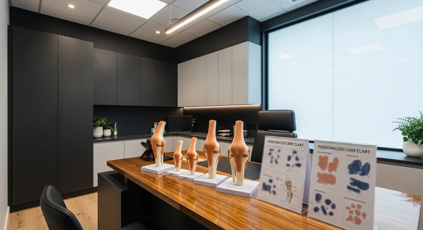
The knee joint is a vital and intricate hinge joint that enables walking, running, and various leg movements. It articulates between three main bones: the femur (thighbone), the tibia (shinbone), and the patella (kneecap). The femur is the longest and strongest bone in the body, with its rounded condyles fitting into the flat top of the tibia. The patella sits in a groove on the anterior side of the femur called the trochlear groove.
This joint's stability and motion are maintained through an extensive network of ligaments, including the anterior cruciate ligament (ACL), posterior cruciate ligament (PCL), and the medial and lateral collateral ligaments. These structures prevent excessive movement and provide support during activity.
Surrounding the bones are articular surfaces covered by smooth hyaline cartilage, which reduces friction and absorbs shock during movement. The surfaces of the tibia and femur are further stabilized by menisci—two crescent-shaped fibrocartilaginous disks—that distribute load and act as shock absorbers, protecting the joint from wear.
Tendons connect the muscles to bones, with the quadriceps tendon attaching the quadriceps muscle at the front of the thigh to the patella, while the patellar tendon connects the patella to the tibia. These facilitate joint movements such as extension and flexion.
Overall, the knee's complex anatomy allows it to bear weight, support the body, and perform a wide range of motions with stability and minimal friction.
Major Ligaments and Their Functions

What are the main ligaments of the knee joint?
The knee joint relies on several crucial ligaments that stabilize and control its movement. The primary ligaments include the anterior cruciate ligament (ACL), posterior cruciate ligament (PCL), medial collateral ligament (MCL), and lateral collateral ligament (LCL).
The ACL and PCL are located inside the joint capsule. The ACL prevents the tibia from sliding forward under the femur and plays a key role in controlling rotational movements of the knee. Conversely, the PCL stops the tibia from moving too far backward and stabilizes the knee during hyperflexion.
Surrounding the knee are the MCL and LCL. The MCL is found on the inside (medial side) of the knee and protects against inward forces that could push the knee inward. The LCL is on the outside (lateral side) and resists outward forces, preventing the knee from buckling outward.
Roles of each ligament in stability
These ligaments work together harmoniously to maintain knee stability. During activities such as walking, running, or jumping, they prevent excessive movements that could lead to injury.
The ACL and PCL are essential for maintaining the front-to-back stability of the knee. The MCL and LCL help control side-to-side motions, especially during lateral impacts.
Their importance in movement
Without these ligaments functioning properly, the knee would be prone to dislocation, instability, and damage to other structures like the menisci or articular cartilage.
Effective functioning of these ligaments ensures smooth hinge motions, enabling activities from simple walking to complex athletic moves.
Understanding the structure and function of these ligaments emphasizes their importance in normal knee movement and stability, and highlights why injuries to them can be so debilitating.
For more details on how these ligaments contribute to knee function and how to preserve their health, search for "knee ligaments functions and stability".
Roles of Menisci and Cartilage
 The structures within the knee joint, particularly the articular cartilage and menisci, serve distinct yet complementary functions. Articular cartilage is a smooth, glassy hyaline cartilage that covers the ends of the femur, tibia, and patella. Its primary role is to facilitate smooth movement of these bones against each other, reducing friction as the joint operates. Additionally, articular cartilage acts as a shock absorber, cushioning the impact during activities such as walking, running, or jumping.
In contrast, the menisci are two crescent-shaped fibrocartilaginous discs located between the femur and tibia. Their main functions are to deepen the tibial surface, evenly distribute the load transmitted through the joint, and improve joint stability. Because the menisci conform to the shape of the femoral condyles, they increase contact area, which helps in reducing the pressure exerted on the articular cartilage.
The difference between articular cartilage and menisci lies in their structure and function. Articular cartilage provides a low-friction, smooth surface for movement and absorbs minor shocks. The menisci, on the other hand, are more substantial, acting as shock absorbers and stabilizers, especially during weight-bearing activities.
Damage or deterioration of these structures can lead to problems like osteoarthritis. When articular cartilage wears down, bones may rub directly against each other, causing pain and inflammation. Meniscal tears can disturb load distribution and destabilize the joint, increasing the risk of further cartilage damage.
More info search queries such as “menisci cartilage shock absorption knee” highlight the importance of these structures in maintaining joint health, protecting against wear and tear, and ensuring smooth, pain-free motion.
Muscles, Tendons, and Their Role in Movement
 The knee joint's ability to move efficiently and withstand stress relies heavily on the coordinated action of various muscles and their connecting tendons. Understanding these components is key to appreciating how the knee functions.
The quadriceps muscle group, located at the front of the thigh, plays a vital role in extending the knee. It comprises four main muscles: vastus lateralis, vastus intermedius, vastus medialis, and rectus femoris. These muscles converge into the quadriceps tendon, which attaches to the superior part of the patella.
Conversely, the hamstring muscles at the back of the thigh—including biceps femoris, semimembranosus, and semitendinosus—are responsible for knee flexion. They connect to the tibia and fibula via tendons, facilitating bending motions essential for walking, running, and jumping.
The gastrocnemius, part of the calf, also crosses the knee joint and aids in knee flexion while contributing to plantarflexion of the foot.
Tendons are robust fibrous tissues connecting muscles to bones, crucial for transmitting force during movement. The quadriceps tendons extend from the quadriceps muscles to the patella, effectively translating muscular contractions into straightening of the leg.
The patellar tendon, also called the ligament of the patella, connects the bottom of the patella to the tibial tuberosity. It works together with the quadriceps tendon to facilitate powerful knee extension.
These tendons not only enable movement but also serve as common sites for injuries like tendinitis or tears, especially in athletes.
In sum, the knee's mobility hinges on the harmony between the quadriceps and hamstring muscles and their associated tendons. Their strength and flexibility are fundamental to everyday activities, athletic performance, and knee joint stability.
For a detailed look into the anatomical structure and interconnected functions of the muscles and tendons around the knee, search for "knee muscles tendons anatomy" to find comprehensive diagrams and educational resources.
Knee Injuries and Clinical Considerations
What are common injuries and conditions related to the knee?
The knee is a complex joint prone to a variety of injuries and conditions that can impair function and cause pain. Among the most common are ligament tears, such as those involving the anterior cruciate ligament (ACL), posterior cruciate ligament (PCL), and the collateral ligaments (medial and lateral). These injuries often occur during sports activities or traumatic impacts when the knee is subjected to sudden twisting or direct blows.
Meniscal tears are another widespread problem, typically caused by twisting motions or sudden pivots, especially in athletes. The menisci act as shock absorbers and stabilizers between the femur and tibia; damage here can lead to joint instability and pain. Bone fractures, particularly of the kneecap (patella), can result from high-impact traumas like falls or car accidents.
Dislocations, where the bones slip out of their normal position, are serious injuries requiring immediate medical intervention. These can damage surrounding tissues, including blood vessels and nerves. Beyond structural injuries, conditions like tendinitis—most notably patellar tendinitis—Bursitis involving inflammation of fluid-filled sacs in the joint, and degenerative diseases such as osteoarthritis and rheumatoid arthritis are common causes of chronic knee pain.
Mechanical problems also contribute to knee issues. These include loose bodies within the joint, iliotibial band syndrome, and patellar malalignment. All these issues can lead to swelling, pain, limited mobility, and instability, affecting everyday activities.
Signs, symptoms, and management
Patients with knee injuries often present with pain, swelling, and difficulty moving the joint. They may notice a popping or tearing sensation at the time of injury. Instability or a feeling of the knee giving way is common in ligament injuries. Mechanical symptoms such as locking or catching may occur with meniscal tears.
Management strategies depend on the severity and type of injury. Mild strains and strains may be treated with rest, ice, compression, and elevation (RICE), along with physical therapy to strengthen surrounding muscles. More severe injuries, like ligament tears or complex fractures, might require surgical intervention, including arthroscopic repairs or reconstructions. Postoperative rehabilitation is crucial to restore function and strengthen the joint.
Preventative measures include proper training, maintaining flexible and strong muscles around the knee, and using appropriate equipment during sports activities.
Search terms for more information include: knee injuries, common causes, treatments.
Conclusion and Educational Significance
A comprehensive understanding of the knee joint's anatomy underscores its vital role in human mobility and stability. Recognizing the interplay of bones, cartilage, ligaments, muscles, and other structures enhances clinical diagnosis, injury prevention, and effective treatment strategies. As the largest joint in the body, the knee's complex architecture demands detailed knowledge for healthcare professionals, students, and patients alike. Continued education through diagrams, models, and clinical case studies will foster better management and recovery for knee-related conditions, ultimately supporting mobility and quality of life.
References
- Knee anatomy including ligaments, cartilage and meniscus
- The Knee Joint - Articulations - Movements - Injuries
- Knee - Physiopedia
- Knee joint: anatomy, ligaments and movements - Kenhub
- Knee Anatomy - William Robert Volk, MD
- Anatomy of the Knee - Arthritis Foundation
- Understanding Knee Anatomy - How a Knee Works | DePuy Synthes
- Anatomy, Bony Pelvis and Lower Limb, Knee - StatPearls - NCBI
- Knee Anatomy: Muscles, Ligaments, and Cartilage
- How understanding knee anatomy can prevent knee injuries