Understanding vastus lateralis rupture and its clinical implications
Vastus lateralis rupture, a rare yet significant injury, can profoundly impact knee functionality and mobility. As part of the quadriceps muscle group, the vastus lateralis plays a crucial role in extending the knee and stabilizing the patella. Despite its importance, isolated ruptures of this muscle are exceedingly uncommon, which often poses diagnostic challenges. Recognizing the injury’s signs, understanding its anatomy, and choosing appropriate management strategies are essential for optimal recovery. This article delves into the anatomy and function of the vastus lateralis, discusses clinical presentation and diagnostic modalities, explores treatment options, and underscores the importance of tailored rehabilitation.
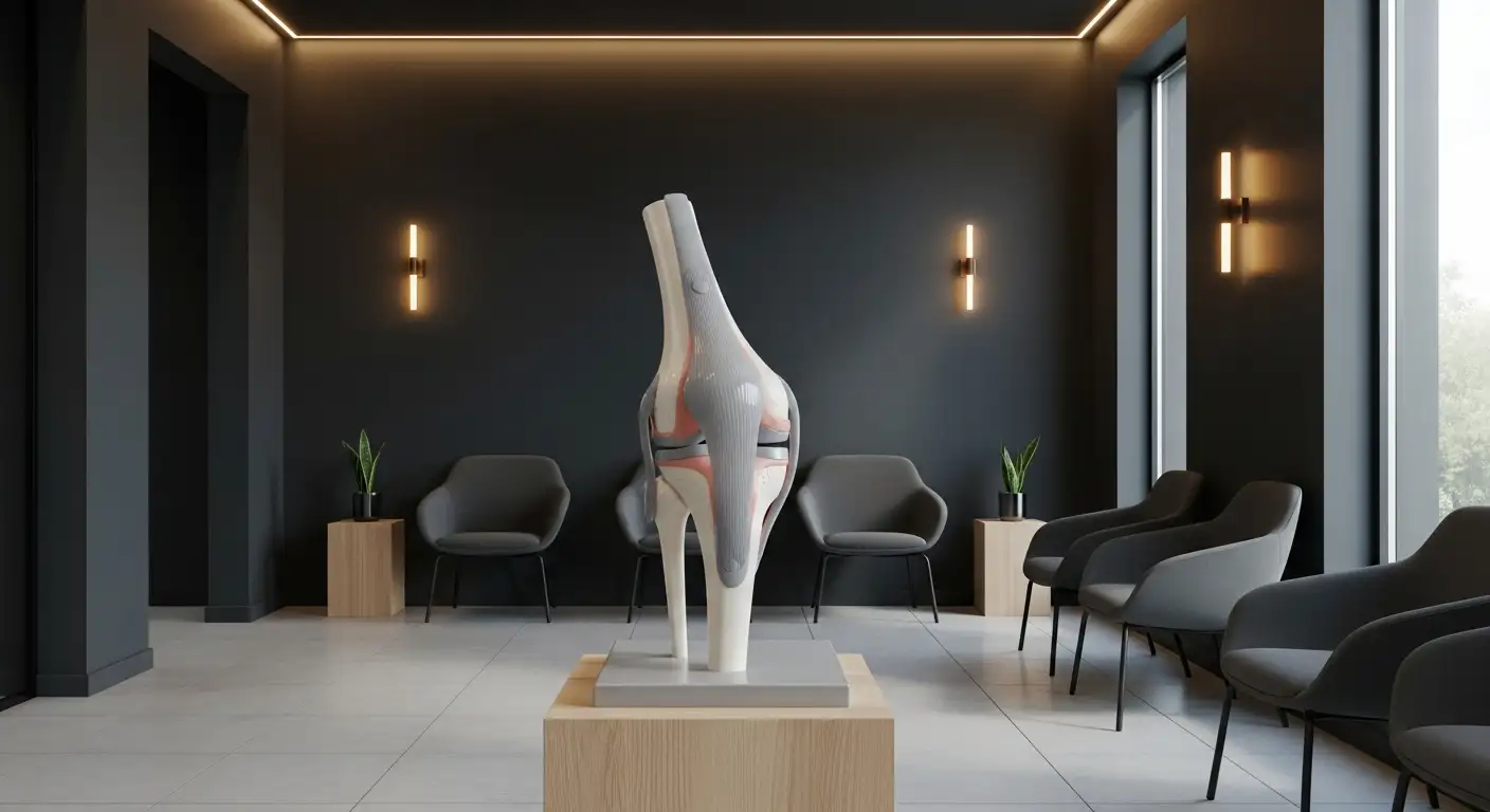
Anatomy and Function of the Vastus Lateralis
What is the anatomy and function of the Vastus Lateralis muscle?
The vastus lateralis is the largest of the four muscles that make up the quadriceps group, situated on the lateral (outer) side of the thigh. Its broad origin includes the intertrochanteric line of the femur, the greater trochanter, the linea aspera, and the lateral intermuscular septum. It extends down the thigh to insert into the lateral border of the patella and the tibial tuberosity through the quadriceps tendon.
This muscle is primarily innervated by the femoral nerve, originating from nerve roots L2 to L4. Its blood supply comes mainly from the lateral circumflex femoral artery, ensuring adequate oxygenation and nutrients for its activity.
Functionally, the vastus lateralis plays a critical role in extending the knee joint, which is essential for movements like walking, running, jumping, and standing up from a seated position. It also contributes to stabilizing the patella, supporting proper tracking during knee movement, and maintaining knee stability during dynamic activities.
Anatomically, the muscle is a unipennate structure covered by fascia lata, and its strength is vital for overall quadriceps functionality. Damage or tearing of this muscle, although rare, can significantly impair knee extension and activity, making its understanding and preservation crucial in sports and trauma contexts.
Symptoms and Clinical Presentation of Vastus Lateralis Rupture
What are the symptoms of a Vastus Lateralis tear?
Symptoms of a Vastus Lateralis tear often appear suddenly during activity or trauma. Patients typically experience an abrupt and sharp pain along the outer thigh or lateral knee. Swelling and tenderness develop rapidly at the injury site, accompanied by discomfort when touching the area.
A distinctive feature is the palpable defect—a depression or gap at the lateral aspect of the thigh or just above the kneecap—indicating a tear in the tendon. Visually, there might be a visible dent or deformity, along with swelling and possible bruising (ecchymosis).
Muscle weakness is common, making it difficult to perform movements like straightening or extending the knee. Patients often report difficulty walking or bearing weight on the affected leg, especially during activities like climbing stairs, running, or jumping.
In some instances, a patient may hear a pop or feel a sudden tearing sensation at the moment of injury. This can be followed by difficulty with knee motion and increased discomfort with movement.
Identifying these symptoms promptly helps healthcare providers diagnose the injury accurately. Proper assessment typically involves physical examination and imaging, such as MRI or ultrasound, to confirm whether the tear is partial or complete.
Overall, these symptoms indicate damage to the tendon or muscle tissue, impairing leg strength, mobility, and functional ability. Early diagnosis and treatment are essential to restore knee function and prevent long-term complications.
Diagnosis: Clinical and Imaging Strategies
How is a Vastus Lateralis rupture diagnosed?
Diagnosing a rupture of the vastus lateralis tendon involves a combination of clinical evaluation and imaging studies. Clinicians begin with a detailed physical examination, looking for signs like localized tenderness along the lateral thigh, swelling, bruising, or a palpable defect near the lateral patellar insertion. Weakness in knee extension or difficulty performing straight leg raises may also be evident, especially in cases of complete rupture.
Due to the rarity and subtlety of isolated vastus lateralis injuries, imaging plays a critical role. Ultrasound is a valuable initial tool; it can detect discontinuity of the tendon fibers or a gap at the attachment site, providing real-time assessment. However, MRI remains the preferred imaging modality, offering detailed visualization of soft tissue structures.
MRI has high sensitivity and specificity for diagnosing tendon injuries. It can clearly delineate partial versus complete tears, show the extent of retraction, and identify associated muscle injuries or other soft tissue abnormalities. Specifically, in cases of isolated vastus lateralis rupture, MRI can confirm the diagnosis amidst the complex anatomy of the quadriceps group.
Plain X-rays generally do not show the tear itself but may reveal indirect signs such as an elevated patella (patella alta) or calcifications. These imaging techniques together ensure accurate diagnosis, which is crucial in planning appropriate treatment—whether conservative management or surgical repair.
In summary, diagnosing a vastus lateralis rupture hinges on an attentive physical exam complemented by ultrasound for initial assessment and MRI for definitive visualization. This comprehensive approach ensures prompt and effective intervention, especially given the injury’s potential to be overlooked due to its rarity.
Treatment options and surgical considerations
What are the common treatment options for Vastus Lateralis tears?
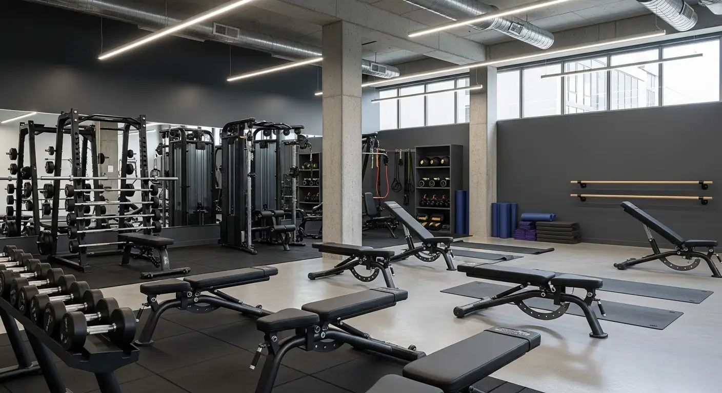
The management of Vastus Lateralis (VL) tendon injuries varies depending on the extent of the tear. For partial tears where the tendon remains mostly intact, conservative approaches are typically effective. These include rest, ice application to reduce swelling, immobilization using braces or supports, and targeted physiotherapy to regain strength and mobility. Such non-surgical treatment aims to allow the tendon to heal naturally while maintaining muscle function.
In cases of complete or full-thickness ruptures, surgical intervention is usually necessary. The primary goal during surgery is to reattach the torn tendon to its original site on the patella. Surgeons often utilize sutures or suture anchors to secure the tissue. Suture anchors are especially useful as they provide firm fixation within the bone, promoting optimal healing.
Indications for surgical repair in complete tears
Surgical repair becomes essential when the tear involves the entire thickness of the tendon, especially if there is significant retraction or if the functional impairment is substantial. Patients presenting with an inability to extend the knee actively, palpable gaps, or confirmed full rupture on MRI are prime candidates for operative intervention.
Early surgical repair, ideally within a few weeks post-injury, dramatically improves functional outcomes. Delayed repairs may be more challenging due to scar tissue formation, and they carry a higher risk of incomplete healing or complications.
Common surgical techniques
Several surgical methods are employed to repair Vastus Lateralis ruptures. The most common techniques involve:
| Technique | Description | Advantages |
|---|---|---|
| Suture anchor fixation | Using anchors embedded into the patella to secure sutures attached to the tendon | Strong fixation, minimal bone damage |
| Transosseous sutures | Passing sutures through drilled holes in the patella | Cost-effective, reliable |
| Graft augmentation | Using autografts or allografts to reinforce the repair in chronic or large tears | Provides additional strength |
The choice depends on factors like tear size, quality of tissue, and surgeon preference.
Postoperative care and immobilization
Post-surgery, immobilization of the knee in full extension generally lasts for about 4-6 weeks to facilitate tendon healing. A hinge knee brace is often used to limit movement while allowing controlled range of motion during rehabilitative phases.
Initial periods involve restricted weight-bearing, progressing gradually to full weight-bearing as tolerated. Physiotherapy plays a vital role, starting with gentle range of motion exercises, advancing to muscle strengthening, proprioception, and functional training.
Most patients can expect to regain near-normal range of motion and strength within 4 to 6 months post-treatment. Full recovery and return to pre-injury activity levels are typically achievable, especially with early intervention and diligent rehabilitation.
Recovery and Rehabilitation After Vastus Lateralis Rupture
What is the recovery process like after a Vastus Lateralis rupture?
Recovering from a Vastus Lateralis rupture depends on the severity of the injury. The initial phase focuses on reducing pain and swelling through rest, ice application, compression, and elevation (RICE). For minor strains (Grade 1), patients often see improvement within 1-2 weeks with proper care and gentle rehabilitation.
For more severe strains or partial tears, especially Grade 2 or 3, recovery may take longer—typically over a month. Physical therapy plays a vital role in restoring muscle strength and flexibility. This begins with gentle range-of-motion exercises, progressing gradually to targeted strengthening activities such as isometric and isotonic exercises.
In cases of full rupture, surgical repair may be required. Postoperative rehabilitation involves immobilization followed by gradual mobilization and physiotherapy, which can take anywhere from 4 to 12 months for full recovery.
Throughout the recovery journey, pain subsides, and swelling diminishes. Patients become able to perform daily activities and more demanding physical tasks once they regain adequate muscle strength, painless range of motion, and functional capacity. Attention to proper rehabilitation protocols helps prevent re-injury and ensures a full return to activity levels, including sports or strenuous exercises.
Challenges in Diagnosis and Treatment
What are the common challenges and considerations in diagnosing and treating Vastus Lateralis injuries?
Isolated ruptures of the vastus lateralis are extremely uncommon, which poses significant hurdles in diagnosis. Since most quadriceps injuries involve multiple muscles, clinicians might overlook an injury isolated to the vastus lateralis or mistake it for more common quadriceps tears. Symptoms such as pain and swelling can mimic other thigh or lateral knee injuries, leading to misdiagnosis.
Accurate diagnosis often requires high suspicion, especially following trauma or overuse activities like weightlifting or sports. Physical examination may reveal tenderness and a palpable defect, but these are not specific. Imaging becomes crucial to confirm the injury. MRI is the gold standard for visualizing soft tissue injuries, distinguishing partial from complete ruptures, and assessing the extent of retraction.
Ultrasound can also be useful and more accessible in some settings, providing dynamic assessment of the tendon.
Deciding on the right approach—surgical versus conservative—depends on the tear’s severity, location, patient activity level, and functional impairment. Complete ruptures generally require surgical repair for optimal recovery, while partial tears may heal with conservative measures like physiotherapy and immobilization.
Given the rarity of isolated vastus lateralis ruptures, healthcare providers need to maintain a high index of suspicion. Incorporating advanced imaging techniques early can prevent missed diagnoses and guide appropriate management.
Overall, these injuries demand careful assessment, timely imaging, and individualized treatment plans to ensure favorable outcomes. Increased awareness is essential to improve diagnosis and optimize recovery for such rare cases.
Case Studies and Clinical Insights
Case example of a 50-year-old male weightlifter
A notable case involves a 50-year-old recreational weightlifter who experienced an isolated rupture of the vastus lateralis during deadlifting. The injury was sudden, characterized by sharp pain on the outer thigh and swelling. MRI imaging confirmed a full-thickness tear of the vastus lateralis with about 3 centimeters of retraction.
Successful surgical repair and return to activity
The patient underwent surgical repair using suture anchors to reattach the torn tendon to the lateral part of the patella. Postoperative care involved immobilization followed by a structured physiotherapy program focusing on gradual range of motion and muscle strengthening. The patient was able to resume full activity and returned to sports and daily activities within 4 months, demonstrating excellent recovery and muscle function.
MRI role in diagnosis
MRI played a crucial role in diagnosing this rare injury, providing detailed visualization of the tendon rupture, extension, and retraction. It differentiated this injury from other lateral thigh problems and guided the surgical plan. MRI remains the preferred imaging modality for such isolated muscle or tendon tears, especially when clinical examination alone is inconclusive.
Implications for clinical practice
This case emphasizes the importance of considering an isolated vastus lateralis rupture when evaluating lateral thigh pain post-injury, even in healthy, active individuals. Early MRI assessment facilitates accurate diagnosis, and surgical repair offers excellent outcomes in complete tears. These insights highlight the need for prompt, precise diagnosis and tailored treatment strategies to ensure patients regain full muscle function and avoid long-term disability.
Advancing Awareness and Management of Vastus Lateralis Injuries
Though exceedingly rare, isolated rupture of the vastus lateralis demands high clinical suspicion and precise diagnostic tools like MRI for accurate identification. Treatment strategies should be tailored to the injury's severity, with conservative management sufficing for partial tears and surgical repair being essential for full-thickness ruptures. Early intervention and dedicated physiotherapy are key to restoring full function and enabling patients to return to their previous activity levels. As clinical cases demonstrate, successful outcomes are achievable with appropriate management, emphasizing the need for awareness of this rare but impactful injury among healthcare providers. Improved understanding can drive better diagnosis, treatment, and ultimately, improved patient quality of life.
References
- Quadriceps Tendon Tear - Physiopedia
- Vastus Lateralis Tendon Rupture & Treatment | Dr HC Chang
- Isolated Vastus Lateralis Rupture and Repair Using Suture Anchor ...
- Quadriceps Tendon Rupture - StatPearls - NCBI Bookshelf
- Isolated Rupture of the Vastus Lateralis Tendon - LWW.com
- Quadriceps Tendon Tear: Symptoms, Tests & Treatment
- Isolated Tear of the Vastus Lateralis Tendon: A Rare Case Managed ...