Introduction to Knee Osteoarthritis and Its Management
Knee osteoarthritis (OA) is a common degenerative joint disease characterized by gradual wear and tear of the articular cartilage, leading to pain, stiffness, and reduced mobility. As the most prevalent form of arthritis affecting the knee, it significantly impacts quality of life, especially among older adults. Orthopedic specialists play a pivotal role in diagnosing and managing this condition through a comprehensive approach that combines clinical assessments, advanced imaging, and tailored treatment strategies. This article explores how orthopedic physicians identify knee osteoarthritis, evaluate its severity, and implement effective interventions aimed at alleviating symptoms and slowing disease progression.
Anatomical and Pathological Foundations of Knee Osteoarthritis
What anatomical features of the knee are relevant when diagnosing osteoarthritis?
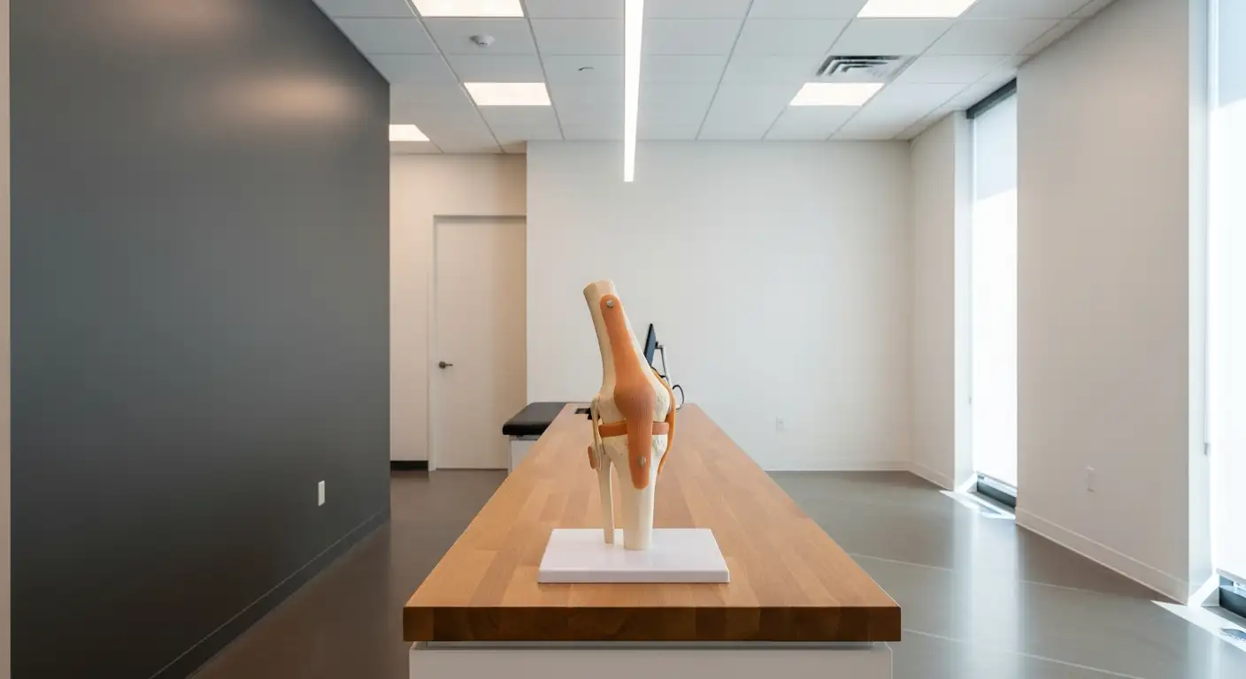
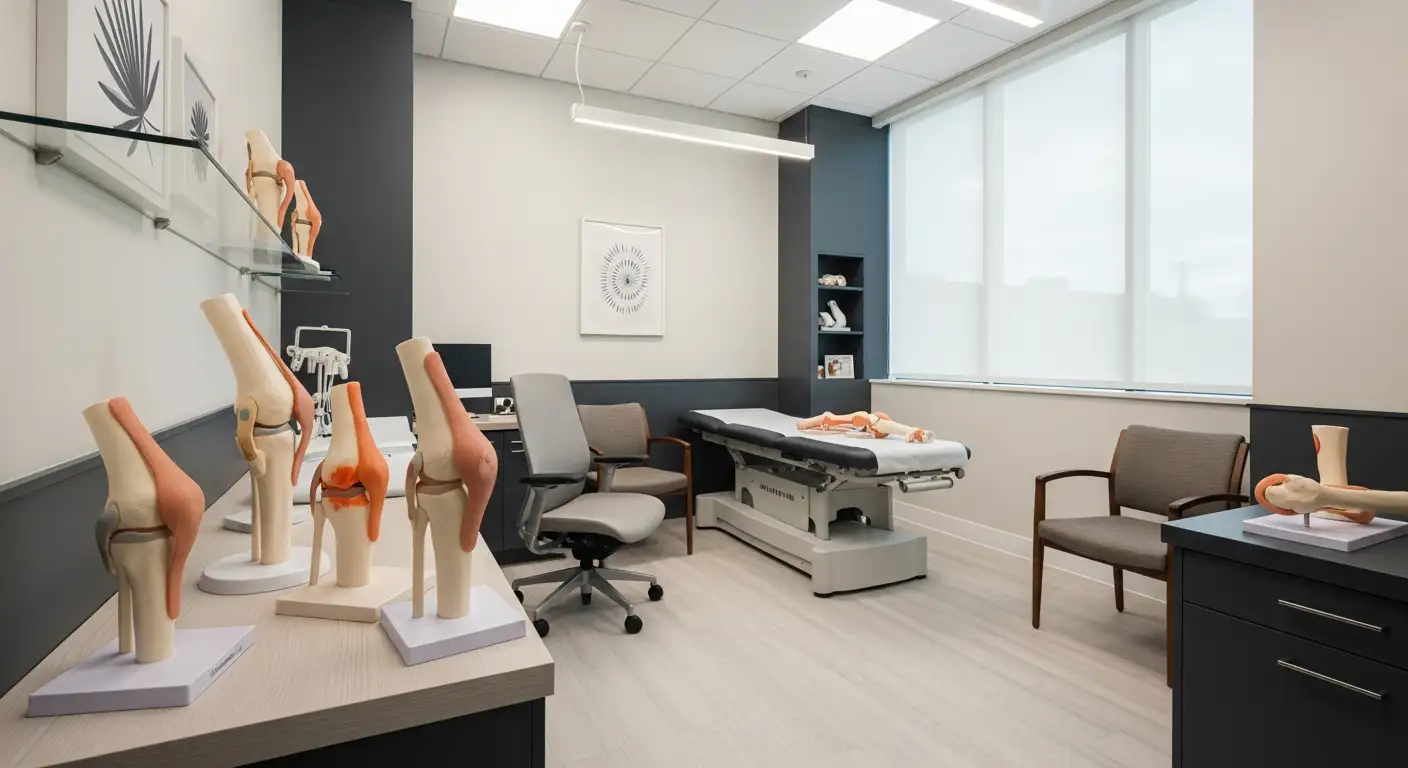
Understanding the anatomy of the knee is essential in diagnosing osteoarthritis accurately. The knee joint is primarily composed of the ends of the femur (thighbone), the tibia (shinbone), and the patella (kneecap). These bones are covered by a smooth layer of articular cartilage, which allows for pain-free movement. The degeneration of this cartilage is a critical hallmark of osteoarthritis.
Surrounding the joint are soft tissues like the menisci, which act as shock absorbers, and collateral ligaments (such as the ACL, PCL, MCL, and LCL), which provide stability. Damage or thinning of the menisci can lead to increased joint stress and faster progression of osteoarthritis.
Bone structures also play a vital role; subchondral bone lies just beneath the cartilage and often shows changes like sclerosis (hardening) or cyst formation in osteoarthritis. Bone spurs, known as osteophytes, develop at joint margins and are visible on X-rays, serving as indicators of the disease.
The synovial membrane, which lines the joint capsule, along with other soft tissues, can become inflamed in some cases, contributing to symptoms. Overall, the complex anatomy of the knee—including bones, cartilage, soft tissues, and ligaments—is integral to both the development and diagnosis of osteoarthritis.
Degenerative changes in joint structures
Osteoarthritis involves progressive deterioration of joint structures. The earliest change is the breakdown of articular cartilage, which loses its smoothness and elasticity, leading to increased friction during joint movement. As cartilage thins and wears away, the underlying subchondral bone becomes exposed.
This exposure results in bone remodeling and the formation of osteophytes or bone spurs, which can be seen on X-rays as bony projections at joint margins. Subchondral sclerosis, characterized by increased bone density beneath the cartilage, occurs as a response to abnormal stress. Additionally, cyst formation within the subchondral bone can occur, further weakening the joint structure.
Surrounding soft tissues may also undergo changes; the synovium can become inflamed (synovitis), and the joint capsule may thicken. These changes contribute to symptoms like swelling, pain, and reduced mobility, marking the progression of osteoarthritis.
Progression of cartilage deterioration and subchondral bone changes
Initially, cartilage degeneration may be mild, with slight surface irregularities. Over time, continued wear leads to significant cartilage loss and exposure of subchondral bone. Bones respond by forming osteophytes at joint surfaces and margins.
Subchondral bone changes accelerate as the joint adapts to abnormal load distribution. The increased bone density (sclerosis) stiffens the joint, reducing its shock-absorbing capacity. In advanced stages, cysts and extensive bone deformities can develop, causing joint instability.
This pathological progression results in characteristic symptoms: persistent pain, stiffness, swelling, and instability, which often worsen with activity and improve with rest. Recognizing these stages through imaging and clinical evaluation helps guide treatment planning.
Overview table of knee osteoarthritis pathology
| Structural Feature | Change in Osteoarthritis | Diagnostic Significance | Associated Symptoms |
|---|---|---|---|
| Articular cartilage | Thinning, erosion, complete loss | Joint space narrowing on X-ray | Joint pain, stiffness, crepitus |
| Subchondral bone | Sclerosis, cyst formation | Detectable via X-ray and MRI | Pain, decreased mobility, swelling |
| Osteophytes | Bone spurs at joint margins | Visible on X-rays | Stiffness, deformity |
| Soft tissues | Synovitis, thickening, inflammation | Ultrasound, MRI | Swelling, warmth, tenderness |
This comprehensive understanding of anatomical and pathological features forms the backbone of diagnosing osteoarthritis, especially in the knee, and aids clinicians in developing tailored treatment strategies.
Diagnostic Approach: Clinical Examination and Patient History

Importance of patient medical history
Orthopedic specialists start diagnosing knee osteoarthritis by obtaining a detailed medical history from the patient. They inquire about the onset and duration of symptoms such as joint pain, stiffness, swelling, or grinding sounds during movement. The doctor also asks about prior knee injuries, surgeries, or other medical conditions that might influence joint health. Understanding lifestyle factors, such as activity levels, weight, and previous treatments, helps form a comprehensive picture.
Physical examination techniques and findings
A thorough physical exam follows, where the specialist assesses the affected knee's condition. They check for signs like tenderness, swelling, warmth, redness, and crepitus (clicking or grinding noise). Range of motion tests reveal stiffness or difficulty in moving the joint. The physician evaluates joint stability and looks for deformities such as bowing or knock-knees. The exam also includes palpation of surrounding tissues to detect additional sources of pain or inflammation.
Assessment of symptoms and functional limitations
The specialist evaluates how symptoms impact daily activities. They note if the patient experiences pain during weight-bearing or activity, morning stiffness lasting less than 30 minutes, or locking and instability episodes. These insights help determine the severity of osteoarthritis and the extent of functional impairment. This assessment guides subsequent diagnostic testing and treatment planning.
How do orthopedic specialists diagnose knee osteoarthritis?
Orthopedic specialists diagnose knee osteoarthritis primarily through a comprehensive clinical assessment that includes reviewing the patient's medical history and symptoms, such as joint pain, stiffness, and functional limitations. They perform a physical examination to evaluate joint tenderness, swelling, range of motion, and signs like crepitus or deformities, along with checking for associated conditions like joint instability. Imaging tests, especially X-rays, are employed to identify characteristic features such as joint space narrowing and bone spurs. MRI or ultrasound may be used for complex cases to assess soft tissues and cartilage. Blood tests and joint fluid analysis help exclude other causes like rheumatoid arthritis or gout but are not definitive for osteoarthritis.
The Role of Imaging and Laboratory Tests in Diagnosis
 When diagnosing knee osteoarthritis, healthcare professionals employ several diagnostic procedures to confirm the condition and assess its severity.
X-rays are the most common imaging test used, as they effectively reveal hallmark signs such as joint space narrowing, the development of bone spurs (osteophytes), and changes in bone shape. These images help confirm cartilage loss and joint deformation. In cases where more detailed images are needed, MRI scans are invaluable. They provide comprehensive visualization of soft tissues, including cartilage, muscles, tendons, and the synovium, allowing early detection of cartilage wear and inflammation that might not be visible on X-rays.
Ultrasound also plays an important role in assessment, especially for soft tissue evaluation. It allows real-time visualization of the synovium, detecting inflammation, excess fluid, and small bone spurs. Computed Tomography (CT) scans are especially useful for detailed bone assessment and are typically used in surgical planning.
Beyond imaging, laboratory tests are conducted mainly to rule out other conditions that may mimic or coexist with osteoarthritis. Blood tests such as ESR and CRP help exclude inflammatory or autoimmune diseases like rheumatoid arthritis. Joint fluid analysis, or arthrocentesis, involves extracting a small amount of fluid from the joint to check for infections or gout, which require different treatments.
In summary, the comprehensive use of imaging—X-rays, MRI, ultrasound, and CT—along with laboratory investigations, provides a detailed understanding of joint health. This combination ensures accurate diagnosis, guides treatment strategies, and helps monitor disease progression.
Clinical Assessment Techniques for Accurate Diagnosis

What are the clinical assessment techniques used by doctors for knee osteoarthritis?
Diagnosing knee osteoarthritis accurately relies heavily on a detailed physical examination combined with imaging studies. Doctors perform a series of specific assessment techniques to evaluate the condition of the joint, identify signs of degeneration, and rule out other causes.
The examination begins with inspection, where the physician looks for visible swelling, deformities, muscle atrophy, and alignment issues such as varus or valgus deformities. Checking for swelling involves observing the joint for puffiness, which may indicate effusion, and inspecting the skin for redness or warmth, signs of inflammation.
Palpation is used to feel for tenderness over the joint surfaces, warmth, and the presence of fluid within the joint (effusion). Tender spots often correlate with areas of cartilage wear or inflammation. The doctor may also check for a bulge or swelling behind the knee, known as a Baker's cyst.
Range of motion testing involves actively asking the patient to move the knee through its normal flexion and extension limits, and then passively moving the joint to assess for restrictions, pain, or crepitus—clicking or grinding noises caused by rough cartilage surfaces.
Joint stability is tested through specific maneuvers like the Lachman or anterior drawer tests—although these are more relevant for ligament injuries, they can also help identify joint laxity or instability, especially if there are associated injuries.
Observation of gait and weight-bearing movements provides vital clues about how the joint performs under normal conditions. Abnormal gait patterns, limping, or difficulty walking may indicate joint pain and functional impairment.
To confirm the clinical suspicion and assess severity, radiographic imaging such as X-rays is employed. X-ray findings often include joint space narrowing, osteophyte (bone spur) formation, subchondral sclerosis, and cysts that support the diagnosis of osteoarthritis.
In conclusion, a combination of inspection, palpation, range of motion testing, stability assessment, and gait analysis forms the foundation of a comprehensive clinical evaluation. These techniques, alongside imaging results, help orthopedic doctors accurately diagnose knee osteoarthritis and tailor effective treatment plans.
Identifying Symptoms and Their Impact on Daily Life
What symptoms are common in knee osteoarthritis?
Knee osteoarthritis often presents with several characteristic symptoms that can interfere with daily activities. Most notably, patients experience joint pain that tends to worsen with activity or after periods of rest. This pain may be dull or sharp and is often accompanied by stiffness, particularly in the morning or after sitting for long periods.
Swelling and tenderness around the knee are also common, reflecting underlying inflammation. Many individuals notice a grinding, cracking, or clicking sensation during movement, a phenomenon known as crepitus, which occurs due to cartilage wear and rough joint surfaces.
As osteoarthritis advances, additional signs may include a decreased range of motion, making it difficult to fully bend or straighten the knee. Some patients observe joint instability, feeling as if the knee might give way unexpectedly. Visible changes can also develop, such as swelling, bone spurs, or deformities like bowing or knock-knees.
Locking episodes, where the knee temporarily sticks in a bent or straight position, may occur due to loose cartilage fragments or joint capsule irregularities. This combination of symptoms tends to develop gradually, intensifying over time, and can significantly impair mobility and overall quality of life.
Understanding these symptoms helps patients recognize early signs of osteoarthritis and seek appropriate evaluation and treatment to manage their condition effectively.
Understanding the Progression and Stages of Disease
 Knee osteoarthritis advances through distinct stages that reflect increasing joint damage and changing symptoms. Recognizing these stages helps in planning appropriate treatment strategies and understanding when surgical options might be necessary.
Initially, the disease is classified as Stage 1, or minor osteoarthritis. At this point, joint damage is minimal, and there are typically no noticeable symptoms. The cartilage, which cushions the ends of bones, begins to show early signs of wear, but patients often experience no pain or restriction in movement.
In Stage 2, or mild osteoarthritis, cartilage deterioration becomes more pronounced. Patients may start to notice occasional pain and stiffness, especially after activity. Small bone spurs, also known as osteophytes, may appear and can be visible on X-ray imaging. During this phase, many individuals can still maintain regular activity levels with the help of conservative treatments.
As osteoarthritis progresses to the moderate stage (Stage 3), cartilage loss is significantly increased. This results in more frequent and intense pain, persistent stiffness, swelling, and a decline in joint function. The joint surfaces become uneven, and larger bone spurs can develop, leading to discomfort and potential difficulty with mobility.
In the most advanced stage, Stage 4 or severe osteoarthritis, cartilage is nearly or completely worn away. Bones are exposed and may grind against each other, causing constant pain and inflammation. This stage is often associated with joint deformity, reduced range of motion, and possible joint instability or immobilization.
The transition from mild to severe disease can occur over several years, but the progression rate varies based on factors like genetics, injury history, body weight, and activity level.
Implications for treatment are closely tied to these stages. Early, minor changes might be managed with lifestyle modification, physical therapy, and medications. More advanced stages often require more aggressive interventions, including injections and ultimately surgery, such as joint replacement, to restore function and reduce pain.
Understanding these stages provides patients and clinicians a roadmap for timely intervention, aiming to slow disease progression and improve quality of life.
Management Strategies and Treatment Options

What are the treatment options for knee osteoarthritis?
Knee osteoarthritis treatment involves a blend of approaches tailored to manage symptoms and slow disease progression. Initially, conservative measures are emphasized, including lifestyle modifications like weight loss and low-impact exercises. These help reduce joint stress and improve mobility.
Physical therapy plays a vital role by strengthening the muscles around the knee, improving flexibility, and reducing pain. Assistive devices such as braces, shoe inserts, or walking aids can provide additional support.
Medications are commonly used, starting with acetaminophen for mild pain. Nonsteroidal anti-inflammatory drugs (NSAIDs) help reduce inflammation and discomfort. In some cases, corticosteroid or hyaluronic acid injections may deliver short-term relief, especially during flare-ups.
Supporting therapies like heat or cold application, acupuncture, and other alternative treatments can also ease symptoms. Patients are encouraged to modify activities to avoid unnecessary joint stress.
When non-surgical measures are inadequate, surgical options become necessary. Procedures such as joint osteotomy, partial or total knee replacement, or joint fusion are considered based on the severity and extent of joint damage. These surgeries aim to remove damaged tissue, realign the joint, or replace the joint surfaces with artificial components.
The goal of each intervention is to significantly diminish pain, restore joint function, and improve overall quality of life. An orthopedic specialist helps develop an individualized plan, considering all these approaches to ensure optimal outcomes.
Concluding Insights on Knee Osteoarthritis Management
Accurate diagnosis and a personalized treatment approach are critical in managing knee osteoarthritis effectively. Orthopedic specialists utilize a combination of patient history, physical examination, imaging, and laboratory assessments to confirm diagnosis and determine disease severity. Management focuses on alleviating pain, restoring function, and preventing further joint deterioration through lifestyle modifications, medications, targeted therapies, and surgical options when necessary. Advances in imaging and minimally invasive procedures continue to enhance outcomes, offering hope for improved quality of life for individuals affected by this widespread condition.
References
- Osteoarthritis - Diagnosis & treatment - Mayo Clinic
- How Orthopedic Doctors Help Osteoarthritis
- How Can Orthopedic Physicians Help With Osteoarthritis Treatment?
- Diagnosing Osteoarthritis of the Knee - NYU Langone Health
- Knee Osteoarthritis: Symptoms, Diagnosis, and Treatment Options
- Knee Osteoarthritis: Symptoms, Stages, Causes & Treatment
- Osteoarthritis: Diagnosis, Treatment, and Steps to Take
- The Role of an Orthopedic Doctor in Osteoarthritis Diagnosis and Care
- Knee Arthritis Treatment Scottsdale AZ - Dr Lige Kaplan
- Osteoarthritis: Diagnosis and Treatment - AAFP